

Zoanthid-Eating Nudibranchs
Zoanthid-feeding aeolid nudibranchs
Identification, coral hosts, eggs, treatment and prevention
Written by Christian, Signature FragsLast reviewed:
What Are They
Zoanthid-eating nudibranchs are small, shell-less sea slugs that feed on zoantharian polyps. A confirmed feeding animal warrants prompt isolation of the affected coral, physical removal of pests and eggs, and repeated inspection before any coral is moved or traded.
The hobby name describes a feeding problem, not a single species. Baeolidia rieae has been identified feeding on Zoanthus in aquariums using morphology and DNA. Palythoa-feeding Aeolidiopsis species are also documented.
This guide separates those records from practical husbandry advice. Its removal and monitoring steps are a conservative management framework; they are not a laboratory-tested eradication programme for every species sold under the name “zoa-eating nudi”.
Which Corals Can They Affect
| Coral group | Evidence | Practical meaning |
|---|---|---|
| Zoanthus | Baeolidia rieae feeding record from aquariums. | Inspect suspect colonies and all recently connected zoanthid material. |
| Palythoa | Aeolidiopsis ransoni and related feeders are documented. | Do not assume that a coral sold as a paly is immune. |
| Other corals | This guide does not establish the host range of an unidentified animal. | A slug found on SPS or LPS needs its own identification and feeding evidence. |
Trade names and colour morphs do not determine a nudibranch’s host range. Record the actual coral where possible. An animal on a colony may be travelling across it; photograph repeated association or feeding damage before assigning a species or a prey preference.
A coral that currently looks unaffected can still carry hidden material. Give particular attention to nearby zoanthids, shared rocks, plugs and corals moved from the same source.
Identification
Look under white light for a crawling slug with paired head appendages and rows of fleshy projections along its back. These projections are called cerata. Look for the outline and movement of an animal rather than relying on colour alone.
The research specimens of Baeolidia rieae were small and greenish-brown with broad cerata; a photographed specimen measured 5 mm. Its age was not reported. Photographs of a small specimen should therefore not be treated as proof of a juvenile stage.


- Inspect polyp stalks, the colony mat, crevices, undersides and the junction between coral and plug.
- Use a loupe or macro camera and include a scale in the photograph. Observe against both light and dark backgrounds.
- Watch for independent crawling or cerata moving separately from coral tentacles.
- Do not identify every frilly sea slug as a zoanthid predator. Some aeolids feed on different animals. Seek experienced identification when the host and photograph are inconclusive.
Aeolidiopsis ransoni can be remarkably well hidden on Palythoa. Rudman documented a brown surface-living form and a pale form sheltered beneath the colony surface. A quick look at the top of an open colony may miss a feeding animal.
What Does the Damage Look Like
For practical monitoring, photograph the same area of the colony each day. Persistent local closure, shrinking polyps, damaged skirts or stalks, and actual missing tissue are reasons to investigate. Record whether damage spreads between adjacent polyps or follows a recent change in conditions.

Closed polyps alone do not confirm nudibranchs. Check water stability, sediment, light, flow, stings and recent handling as well. Distinguish intact retracted polyps from lost tissue, and look for other pests such as sundial snails or sea spiders.
Finding a crawling animal alongside recurring damage or a recognised egg mass strengthens the diagnosis. Collect the animal and document it rather than repeatedly disturbing the colony without confirming the cause.
Discovery and Confirmation
Move the suspect colony, submerged, into a dedicated inspection vessel containing matched seawater. Examine all accessible surfaces with magnification. Use gentle water movement to reveal sheltered areas and inspect material that settles in the vessel.
- Collect visible animals with a pipette or fine forceps without squeezing live polyps.
- Photograph the animal, affected coral and any egg ribbon before removal. Record approximate size and location.
- Count what is recovered, then inspect other suspect colonies separately. Avoid mixing new arrivals into the affected group.
- If only a vague shape is visible, maintain isolation and obtain a clearer image or experienced identification.
Handling Zoanthids Safely
Zoanthids and Palythoa require palytoxin precautions. Keep colonies submerged during transfer and work, wear suitable aquarist gloves, eye protection and the respiratory protection recommended by OATA. Keep other people away from the work area. Do not boil, steam, microwave, dry or pressure-spray zoanthid-covered rock. Avoid vigorous brushing and spraying. If exposure is suspected, seek urgent medical attention and explain that palytoxin may be involved.
Eggs
Search for an attached, gelatinous cord or ribbon containing many small pale dots. It may be coiled, folded or partly concealed near the polyps and colony mat. Baeolidia rieae produces a coiled egg cord; photographs of Aeolidiopsis show that a Palythoa feeder’s ribbon can be folded differently.
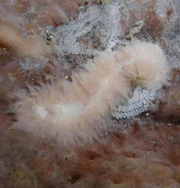

A circular pattern alone cannot identify the parent species. Use the recovered animal, host and egg structure together. Do not label an unrelated snail or Berghia egg mass as a zoanthid pest without supporting evidence.
Remove accessible suspect egg material outside the display while keeping the colony submerged. Lift it with a suitable fine tool into the inspection vessel and recheck the attachment area. Avoid tearing the live mat. If material cannot be reached safely, maintain isolation and seek experienced help.
Treat egg inspection as a separate task from dipping. A dip that dislodges mobile animals does not establish that an attached ribbon has been killed or removed. A clean dip container is therefore not a clearance certificate.
Understanding the Breeding Cycle
Nudibranchs are hermaphrodites, with male and female reproductive organs. Rudman’s account does not support the common claim that nudibranchs reproduce asexually or routinely fertilise themselves. Finding only one animal is still a reason to search carefully for additional animals and eggs.
Across nudibranchs, development may include swimming larvae or direct emergence of crawling young. The route must be established for the species concerned; a timetable from Berghia, Montipora feeders or another nudibranch is not automatically applicable here.
| Stage or question | Evidence and management implication |
|---|---|
| Egg ribbon | Inspect and physically remove accessible material; do not assume a dip has killed it. |
| Hatching and early stages | No verified temperature-specific timetable is provided here for the unidentified aquarium pest. Continue magnified inspections. |
| Sexual maturity | Do not infer age or maturity from size or colour alone. |
| Repeat treatment | Use the current product label and coral tolerance, and record each finding. |
| Host-free period | No universal eradication duration is established here. A calendar deadline alone cannot confirm clearance. |
Coral Rx’s pest article suggests repeating treatment every 4–8 days and extended quarantine. This is manufacturer aquarium guidance, not a controlled, species-specific life-cycle result. It should not be described as proof that all eggs hatch or all young are removed within that interval.
Immediate Containment
Stop selling, trading or transferring affected material while the problem is assessed. Isolate suspect colonies in an independent quarantine system with appropriate temperature, salinity, light and circulation. Another rack in the same connected system does not contain the problem.
- Inspect recently moved corals, shared plugs, rubble and nearby zoanthid colonies. Record their origins and transfer history.
- Use dedicated tools, inspection vessels and rinse containers. Prevent dip or inspection water from returning to any coral system.
- Keep incoming corals separate from the affected group so follow-up findings remain interpretable.
- If colonies are attached to a large rock, plan safe submerged removal. Do not use heating, drying or destructive brushing as a shortcut.
Treatment
Physical Removal
Remove visible animals and accessible egg material in a separate inspection vessel. Recheck the underside, polyp bases and attachment point after removal. Work patiently under magnification and avoid crushing or puncturing polyps.
If a plug or other attachment can be replaced safely without injuring the coral, inspect the removed piece separately and keep it out of clean systems. Cutting away all live tissue or aggressively scraping a zoanthid mat is not a routine substitute for careful inspection.
Coral Dips as a Supporting Measure
Choose a coral dip whose current instructions allow use on the coral concerned. Follow the directions for the exact formulation, measured water volume and exposure time. Do not increase concentration or duration because nothing has fallen off, and do not substitute a household chemical or pesticide recipe.
Coral Rx’s general directions specify separate dip and seawater rinse containers, close inspection and subsequent quarantine. The standard, Pro and Industrial products have different dosing instructions. Use the current bottle’s directions; never dose Coral Rx into the display or quarantine system, and never reuse mixed dip water.
Match the inspection and rinse water to the coral’s temperature and salinity. Check between polyps during treatment, collect visible animals directly and inspect for eggs afterwards. Return the coral to independent quarantine after rinsing.
Record the product, measured dose, duration, animals and eggs found, and the coral’s response. The useful outcome is a documented reduction in findings with recovering coral, not merely completion of a dip.
When Treatment Is Not Working
New animals, fresh egg material or continued tissue loss require another inspection of both the colony and its surroundings. Check for overlooked attachment surfaces, connected suspect corals and a second cause of decline. Reconsider the identification before intensifying treatment.
A colony that cannot be inspected or treated without serious damage may need experienced help or controlled disposal. Follow the zoanthid handling and disposal guidance in reference 8. Do not release pests, coral fragments or aquarium water outdoors or into natural waterways.
Biological Pressure
Coral Rx suggests wrasses as an aid to control. This is supporting aquarium guidance, not proof that a particular fish will remove every hidden animal or egg.
Do not introduce a fish solely as an eradication guarantee. Assess the aquarium’s space, other livestock and long-term husbandry needs. Continue direct removal, egg inspection and quarantine even if fish appear to pick at mobile pests.
Repeated Inspection and Follow Up
Inspect isolated colonies regularly under consistent white light. Daily visual checks are a useful husbandry routine during an active problem; they are not a measured hatching interval. Use magnification for the mat, undersides and plug attachment.
- Keep dated photographs of the same polyps and mark newly damaged areas.
- Record each mobile animal or egg ribbon, including findings from inspection and dip vessels.
- Check how the coral responds to handling before repeating an intervention.
- If a new finding appears, extend monitoring and investigate where it could have been missed or reintroduced.
Quarantine
Quarantine must be physically independent, with suitable husbandry rather than simply a holding container. Use dedicated equipment and avoid sharing water. Keep the affected batch identifiable throughout observation.
A long quarantine period is helpful only when it includes inspection and appropriate action. Repeated unexplained closure or fresh tissue loss needs investigation even if no animal has yet been photographed.
Do not set a universal clearance date from an unconfirmed species name. The duration should reflect the actual findings, treatment response and confidence of inspection. Coral Rx recommends lengthy observation, but that recommendation does not prove eradication at a fixed date.
Final Clearance and Reintroduction
Before reintroduction, review the full observation record and inspect every accessible surface again. Arrange a final confirmation dip where suitable for the coral and the product. Negative findings reduce concern but do not prove that hidden eggs are absent.
- No fresh damage, visible crawling pests or suspect egg material during repeated inspections.
- Recovery of normal polyp appearance and extension, judged alongside stable water conditions.
- Inspection of the underside, colony mat, attachment point and any rock or plug that will be transferred.
- Clean equipment and a transfer route that does not bring old inspection, dip or rinse water into the destination.
If any of those checks remains unresolved, retain the colony in quarantine. Continue observation after reintroduction and keep a record of where the coral was placed.
Recovery
Recovery depends on the tissue left, handling stress and aquarium conditions. Give the colony stable, appropriate light and circulation, avoid repeated relocation and monitor the same polyps over time.
Healthy extension may return before new tissue or additional polyps are apparent. Do not compensate for damaged coral by abruptly increasing food, light or dosing. Base any changes on the system’s measured needs.
Prevention
- Buy from reputable aquaculture sources with clear inspection and quarantine practices.
- Inspect new zoanthids under white light before introducing them to a clean system.
- Use a suitable coral dip as one part of the process, followed by independent quarantine and repeated inspection.
- Examine plugs, rock, colony mats and undersides for eggs as well as mobile animals.
- Keep equipment and water from incoming or affected batches separate from cleared coral.
Source history helps assess risk, but a seller’s assurances or one clean dip cannot replace inspection. Avoid passing suspect material to another reefer while the problem remains unresolved.
The Signature Frags Perspective
Our approach is to make the cause visible, contain the affected material and keep a useful record. Removing the animals you can see is only the beginning when an attached egg ribbon may remain. A deliberate inspection and quarantine process is more informative than repeatedly changing treatments without recording the result.
Christian’s Take
A zoanthid colony that keeps closing deserves a proper look under white light. Once you find a feeding nudibranch, deal with the whole affected batch and inspect the eggs separately. Be patient with the coral, record what you remove and keep the quarantine process going until you have a convincing history of clean inspections.
References
Research, original observations and manufacturer instructions are distinguished below. Practical management steps are conservative husbandry recommendations. Sources checked 4 October 2026.
Cabrito A, Galià-Camps C and Ballesteros M (2022). The risk of the nudibranch Baeolidia rieae in the international aquarium trade. Cahiers de Biologie Marine 63, 1–11. Aquarium host, morphology and spawn observations.
Rudman WB (2002). Aeolidiopsis ransoni Biology. Sea Slug Forum, Australian Museum. Palythoa feeding and camouflage.
Smith SA and Rudman WB (2007). Aeolidiopsis sp in aquarium. Sea Slug Forum, Australian Museum. Original aquarium observation with qualified identification.
Masayoshi N and Rudman WB (2002). Egg ribbon of Japanese Aeolidiopsis. Sea Slug Forum, Australian Museum. Egg-laying photograph and identification discussion.
Rudman WB (1999). Nudibranch questions. Sea Slug Forum, Australian Museum. Hermaphroditism and limits of self-fertilisation evidence.
Coral Rx (2023). Zoanthid Eating Nudibranchs. Manufacturer aquarium advice. Its asexual-reproduction statement is not adopted in this guide.
Coral Rx (2023). Effectively Dip Corals in Coral Rx. General manufacturer instructions; check the current product label.
Ornamental Aquatic Trade Association. How to avoid palytoxin poisoning if you keep corals. Handling, protective equipment, disposal and exposure guidance.
Department of Biodiversity Conservation and Attractions, Western Australia. Nudibranches sea slugs. General reproduction and development; not a zoanthid-pest timetable.
Masayoshi N and Rudman WB (2002). Aeolidiopsis ransoni from Hachijo Japan 2. Sea Slug Forum, Australian Museum. Pale-form and egg-mass photographs; original identification qualified.
Image Credits
Background supplied by Signature Frags and reused from the established guide layout.
Research photographs: Andrea Cabrito, Carles Galià-Camps and Manuel Ballesteros (2022), The risk of the nudibranch Baeolidia rieae in the international aquarium trade, Figure 1B, D and E. Sourced from the University of Barcelona repository. Cropped to the original panels; panel letters and animal content retained. © CNRS – Station Biologique de Roscoff – ABiMS, 2022. Open access does not establish permission for commercial website image reuse. Included in this unpublished draft for review; publication permission remains unconfirmed.
Egg-laying and egg-mass photographs: M. Nishina (Nishina Masayoshi), Sea Slug Forum, Australian Museum (2002), Egg ribbon of Japanese Aeolidiopsis and Aeolidiopsis ransoni from Hachijo Japan 2. Full original photographs retained. The pale-form identification was qualified by the source. Website publication permission remains unconfirmed.
The 5 mm research specimen is labelled with its reported size; its age is unknown. No photograph here is labelled as a confirmed newly hatched zoanthid-eating nudibranch.
More from the Coral Pest Library
Montipora-Eating Nudibranchs →
Goniopora-Eating Nudibranchs →
Explore Pests & Problems