

Goniopora-Eating Nudibranchs
Phestilla goniophaga
Identification, breeding cycle, treatment and prevention
Written by Christian, Signature FragsLast reviewed:
What Are They?
Goniopora-eating nudibranchs are coral-feeding sea slugs. The named species addressed in this guide is Phestilla goniophaga, described in 2020. It consumes living coral tissue and belongs in the coral pest library; “corallivorous predator” is more precise than treating it as an infectious disease.
The practical priorities are to confirm the animal, isolate affected corals, remove mobile pests and egg masses, and keep inspecting. A reassuring dip result is not sufficient evidence to move a questionable coral back into an established collection.
This guide separates identification and life-history research from a conservative husbandry response. Advice for Montipora-eating nudibranchs must not automatically become the treatment protocol for Goniopora.
The original Hong Kong observations record feeding on G. columna, G. djiboutiensis and G. lobata. A separate 2024 survey recorded an unnamed Goniopora-associated Phestilla on G. fruticosa and G. stokesi in Thailand. That record should not automatically be relabelled P. goniophaga.
Identification

Published specimens reached 30 mm; smaller specimens were 8–15 mm. The brownish or pale reddish body bears many elongated cerata that resemble coral tentacles, with a rounded white hump mimicking the polyp mouth. Juveniles have fewer, less-developed cerata. Animals and eggs are commonly beneath colonies.
Use neutral white light rather than relying on the appearance under strong blue lighting. Inspect in a shallow container of matched seawater and keep living tissue submerged while checking. Magnification and a macro photograph can help distinguish an independently moving animal from a retracted or damaged polyp. These are inspection recommendations, not diagnostic tests.
- Inspect the complete colony, including its underside, exposed skeleton, plug and attachment points.
- Look for movement of a separate body, rather than judging colour alone.
- Photograph a suspected animal from above and the side, ideally with a scale beside it.
- Record the host coral and where the animal was found; retain a safely contained specimen for expert identification if necessary.
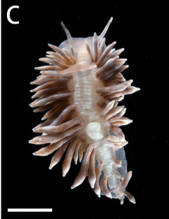
Juveniles — the crawling stage
A documented juvenile with fewer, less-developed cerata. This is a crawling juvenile, rather than the shelled swimming veliger. The original Figure 2C scale is 5 mm.
What Does the Damage Look Like?
A 2023 laboratory study measured size-dependent feeding by P. goniophaga at 1.44–8.33 Goniopora polyps per individual per day. This is an experimental range, not a prediction of how quickly a particular aquarium colony will die.
For practical monitoring, photograph areas with missing tissue or exposed skeleton and compare the same area over successive inspections. Reduced extension, a patch of missing polyps or declining condition should prompt investigation, but none of these observations alone identifies a nudibranch.
- Check whether tissue loss progresses despite stable aquarium conditions.
- Inspect beside newly exposed skeleton and at the colony’s sheltered margins.
- Separate actual tissue loss from polyps that remain intact but retracted.
- Consider recent handling, stings, sediment, unsuitable flow, water instability and other disease processes as competing explanations.
Do not label every declining Goniopora as infested. Confirmation should come from an animal consistent with the identification evidence, or a well-documented egg mass examined in context.
Discovery and Identification
If a pest is suspected, move the coral to a separate inspection vessel rather than trying to blast material off inside the display. Use gentle flow to reveal sheltered surfaces; avoid tearing tissue or forcing the animal back into a crevice. Remove suspicious mobile animals with a pipette or suitable fine forceps where accessible.
Examine the inspection water against both light and dark backgrounds. A phone photograph may help, but a very small animal should not be dismissed simply because its features cannot be resolved. Obtain experienced identification before making a firm species claim.
Distinguish adults, settled juveniles and planktonic larvae. A photograph of a shelled veliger is not a photograph of a miniature crawling adult. The original paper’s Figure 2 identifies those stages separately.
Eggs
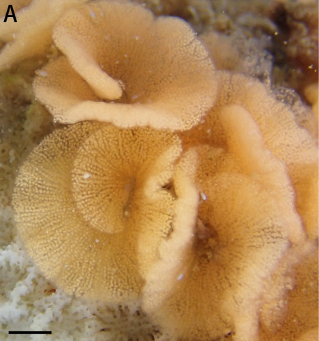
The described egg masses are bright orange coiled ribbons, roughly 1 cm across, firmly attached to coral skeleton. A mass can contain more than 6,000 eggs; researchers observed as many as 20 masses on one colony during the productive season. Embryos developed into swimming veligers in approximately five days. These are observations under the study conditions, not an aquarium guarantee.
As a practical containment measure, inspect sheltered skeleton carefully and physically remove any suspected ribbons while the coral is outside the display. Collect removed material and inspection water for disposal; do not return it to a connected system. Take photographs before removal if identification is uncertain.
Do not assume that removing adults, a clear dip container, or a change in egg colour proves the eggs are dead. The sources reviewed do not demonstrate an egg-killing dip protocol for P. goniophaga.
Understanding the Breeding Cycle
The useful distinction is between development inside the egg mass, hatching, larval competence, settlement and later sexual maturity. “Five days” is not a complete egg-to-egg generation time.
Earlier Guam experiments used the informal name Phestilla sp. 2, which the 2020 description linked genetically to P. goniophaga. In the 2007 experiments, embryos were typically fully developed after about five days and egg masses were opened manually. Cultures were held at approximately 28–32°C. Significant metamorphosis in response to the preferred host was recorded five days after hatching. These separate intervals must not be collapsed into one universal five-day life cycle.
A 2009 study found that waterborne cues from Goniopora could induce metamorphosis, with the strongest response to G. fruticosa in the tested conditions. Settlement responses and adult feeding preferences are related but do not match perfectly.
The 2023 paper reports a shorter 1–3-day period to metamorphic competence across the three studied species. Its accessible abstract does not provide the complete species-specific experimental conditions needed to turn that range into an aquarium treatment timetable.
| Stage | Evidence available | Practical meaning |
|---|---|---|
| Egg development | See the study-specific observations above. | Inspect eggs immediately; do not wait for a planned weekly check. |
| Swimming larvae | A planktonic stage is documented. | Treat connected water as a potential spread route; use independent quarantine. |
| Settlement | Host cues can induce metamorphosis. | Moving a coral to another part of the same system does not isolate it. |
| Juvenile to breeding adult | No reliable aquarium interval established in the reviewed evidence. | Do not invent a maturity deadline or stop treatment on that basis. |
| Complete clearance | No validated aquarium eradication endpoint identified. | Use repeated negative inspections and continued observation, with uncertainty retained. |
Immediate Containment
The following is a conservative biosecurity response inferred from the pest’s host association and larval stage, rather than a tested eradication trial.
- Pause transfers, sales and additions involving the affected group until the extent of the problem is understood.
- Inspect every Goniopora sharing the system, including corals that still look healthy.
- Place suspect corals in a suitable, independently filtered quarantine system with matched conditions. A sump, rack or breeder box sharing display water is not independent quarantine.
- Keep tools, containers and rinse water separate. Clean equipment appropriately before reuse elsewhere.
- Record the last introductions and any recent transfers so exposed corals can be traced.
Quarantine must support the coral’s condition: avoid replacing pest risk with unstable temperature, salinity, oxygenation or inadequate flow. Handle the coral gently and retain the ability to inspect its base.
Treatment
Physical removal
Remove accessible animals during inspection and separately remove suspected egg material from exposed skeleton. Work carefully around the tissue margin. Do not scrape living flesh simply to reach a crevice, and do not automatically frag the whole colony as a first response.
If an inaccessible mounting surface remains suspect, consider replacing a removable plug or cutting away non-living attachment material only when that can be done without disproportionate damage. Destructive rescue fragging requires case-specific judgement, and every retained piece remains under quarantine.
Coral dips as a supporting measure
A commercial coral dip may assist inspection and removal of mobile organisms, but efficacy, coral tolerance and egg killing cannot be assumed for this species. Select a product explicitly suitable for the coral concerned and follow its current instructions in a separate container. Rinse as directed with clean matched seawater; inspect before returning the coral to quarantine.
There is no species-specific dip concentration, contact time or repeat interval established by the research reviewed for this guide. Do not copy an Acropora or Montipora protocol, increase concentrations, mix dip chemicals or combine treatments to compensate for that uncertainty.
Repeated inspection rather than a promised chemical cure
Inspect daily while animals, egg material or progressive damage are being found, using brief checks that limit handling. Remove new findings promptly. Full dips should be based on demonstrated need, coral condition and product guidance; daily observation does not mean daily chemical dipping.
Use a simple log: date, animal count, suspected eggs, location of new damage, removal or treatment performed, and the coral’s response. A recurring finding means the containment and removal process is still incomplete.
Whole system treatments and experimental chemicals
The reviewed primary literature does not validate a reef-safe whole-tank cure for P. goniophaga. Chemical recipes circulated for other pests are not established treatments for this animal. Freshwater, concentrated oxidants and veterinary medicines should not be presented as routine solutions without evidence for both pest efficacy and Goniopora tolerance.
Biological Pressure
Do not buy a fish or invertebrate on the assumption that it will clear this pest. No dependable aquarium predator treatment for P. goniophaga was established in the primary sources reviewed. Existing compatible predators may contribute background pressure, but they cannot replace inspection, egg removal and quarantine.
A lack of visible animals after introducing a predator is not proof of eradication. Sheltered animals and unexamined surfaces still require assessment.
Follow Up
Keep affected corals isolated while findings continue. Review the inspection log for fresh damage, animals or suspected egg material rather than judging success solely by improved polyp extension.
- If a new animal or egg mass is found, remove it and review all exposed corals and equipment again.
- If damage continues without a visible pest, reassess identification and alternative causes rather than escalating chemicals blindly.
- After repeated clean checks, reduce unnecessary handling but continue close observation.
There is no proven number of clean inspections or minimum observation period that guarantees clearance. An inspection programme lasting only until the first clear dip is inadequate evidence for release.
Quarantine and Host Removal
Use quarantine to make inspection repeatable and prevent further transfers. Maintain clear separation between incoming, exposed and cleared corals. New introductions into the same quarantine group can make earlier negative observations less meaningful.
Host removal may be considered as part of a coordinated response, but the published material reviewed does not establish a safe host-free interval for an aquarium. Do not advertise a four-week, six-week or other calendar period as proven to starve this species out.
Removing the visibly affected colony alone may leave other exposed Goniopora in the system. Equally, placing suspect coral back in the display to “test whether anything remains” risks restarting the problem.
Final Clearance and Reintroduction
Before any transfer, complete a deliberate inspection of the whole coral, base and attachment surfaces. Look for mobile animals, suspicious egg material and unexplained fresh tissue loss. Review whether exposed neighbouring corals have also remained free of findings.
A final supporting dip can provide additional inspection information if appropriate for the coral, but a clean dip alone does not rule out hidden eggs or larvae. Reintroduction should be a cautious husbandry decision supported by repeated negative observations; it should not be described as scientifically certified pest-free clearance.
Coral Recovery
Once findings and fresh damage cease, prioritise stable husbandry and limit further disturbance. Monitor salinity, temperature, alkalinity, calcium and magnesium, along with suitable light and flow. Keep exposed areas free of settled debris without directing a harsh jet at compromised tissue.
Feed appropriately for the coral’s condition and avoid sudden changes intended to force rapid recovery. Photograph the same areas to distinguish regrowth from simple changes in polyp extension. Persisting recession or deterioration needs reassessment of water conditions, physical injury and other disease processes.
Prevention
Choose sources with clear inspection and quarantine practices. Inspect every incoming Goniopora, including aquacultured pieces; tank-grown origin does not by itself prove freedom from hitchhikers.
Check the underside and mounting surfaces before introduction and throughout quarantine. Document unusual findings, keep equipment separate, and avoid admitting a coral simply because one inspection or dip was reassuring.
The 2023 experiments support a strong association with Goniopora, but they do not establish that every traded species or colour morph has been tested. As a practical precaution, include the entire exposed Goniopora group in surveillance. Do not automatically classify Alveopora as a confirmed host without identification or specific evidence.
Signature Frags Perspective
The strongest message for this guide is methodical biosecurity: careful sourcing, close inspection, separate quarantine and repeat observation. Treatment should target what is actually found while protecting the coral’s condition. Avoid promising that any single dip or fixed calendar schedule guarantees clearance.
Christian’s Take
With a Goniopora that starts losing tissue, I’d want to know why before reaching for a bottle. Look underneath it, inspect the base and check the rest of the Goniopora in the system. If you find a nudibranch, remove it and look properly for eggs. Then keep checking. The aim is to stop the pest without damaging the coral through repeated heavy-handed treatment. One clean dip is useful information, but it isn’t the finish line. Be methodical and let the coral recover once the feeding has stopped.
References and Evidence Notes
Research checked 3 October 2026. References support the biological claims. Management passages are conservative husbandry recommendations and should not be mistaken for tested species-specific treatment results.
Hu, J., Zhang, Y., Yiu, S.K.F., Xie, J.Y. & Qiu, J.W. (2020). A New Species of Predatory Nudibranch (Gastropoda: Trinchesiidae) of the Scleractinian Coral Goniopora. Zoological Studies 59:62. DOI 10.6620/ZS.2020.59-62. Original description, identification, eggs and genetic connection to earlier Phestilla sp. 2 records.
Yiu, S.K.F. & Qiu, J.W. (2023). Distribution, dietary preference and larval settlement preference of three scleractinain-coral-eating nudibranchs Phestilla spp. (Nudibranchia: Trinchesiidae) from Hong Kong waters. Regional Studies in Marine Science 61:102858. DOI 10.1016/j.rsma.2023.102858. Accessible abstract establishes feeding rate, host association and study-wide competence range; full species-specific timing must not be inferred from the abstract alone.
Ritson-Williams, R., Shjegstad, S.M. & Paul, V.J. (2007). Larval metamorphic competence in four species of Phestilla (Gastropoda; Opisthobranchia). Journal of Experimental Marine Biology and Ecology 351:160–167. DOI 10.1016/j.jembe.2007.06.010. Earlier work on Phestilla sp. 2; distinguishes embryonic development from days after hatching and records warm culture conditions.
Ritson-Williams, R., Shjegstad, S.M. & Paul, V.J. (2009). Larval metamorphosis of Phestilla spp. in response to waterborne cues from corals. Journal of Experimental Marine Biology and Ecology 375:84–88. DOI 10.1016/j.jembe.2009.05.010. Earlier Phestilla sp. 2 experiments examining host cues and differences between settlement response and adult feeding.
Mehrotra, R., Caballer, M., Kaullysing, D., Jualaong, S. & Hoeksema, B.W. (2024). Parasites or predators? Gastropod ectoparasites and their scleractinian host corals at Koh Tao, Gulf of Thailand, with the description of a new species. Symbiosis. DOI 10.1007/s13199-023-00967-z. Field records retain a Goniopora-associated Phestilla as unnamed; identity should not be assumed from host association alone.
Evidence limits that affect the protocol
- No controlled species-specific commercial dip or whole-system eradication trial was identified in this review.
- Juvenile time to sexual maturity and a guaranteed aquarium clearance or host-free period remain unestablished here.
- Reported timings reflect different studies and conditions; they are not a validated timetable at 25°C.
- Symptoms alone do not establish species identity, and an unidentified nudibranch should not automatically be called P. goniophaga.
Image Credits
Background supplied by Signature Frags, reused from the Goniopora & Alveopora care guide.
Adult, juvenile and egg photographs: Juntong Hu, Yanjie Zhang, Sam King Fung Yiu, James Yang Xie & Jian-Wen Qiu (2020), Zoological Studies 59:62, Figures 1A, 2C and 2A. © 2020 Academia Sinica, Taiwan. Original species-description study; figure copies sourced from Reef Builders, Jeremy Gay, 23 June 2023. Cropped to isolate each stage, retaining the original scale bars. No synthetic detail was added.
Image reuse permission has not yet been confirmed. These research photographs are retained in this unpublished draft for review before publication.
More from the Coral Pest Library
Goniopora & Alveopora care guide →
Montipora-eating nudibranch guide →
Drupella coral-eating snails guide →
Explore Pests & Problems